The Origin of Primordial Germ Cells Becoming Caviar — How do potential caviar cells emerge from fertilized eggs in a sturgeon?
Research Press Release | March 04, 2014
-
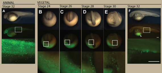 Migration of visualized PGCs in sturgeon embryo. An embryo injected with the GFP-nos3 3′UTR mRNA at the animal pole (A) and at the vegetal pole (B–F). (A) The animal pole labeled embryo at stage 32. (B) The vegetal pole labeled embryo at stage 24. PGCs were initially found around the marginal region of the posterior developing embryonic body at this stage (C) The embryo at stage 26. The PGCs migrated dorsally as the tail rudiment bulged out. Until this stage, the distribution of the labeled PGCs is crescent-like surrounding the developing tail bud. (D) The embryo at stage 28. The PGCs were divided into two populations, at the left- and right-side of the embryonic body. At this stage, fluorescence of PGCs was stronger than during stages 24 and 26. Most PGCs are still localized on the yolk ball. (E) The embryo at stage 30. Most PGCs are located on the yolk extension, but some more ventral were still on the yolk ball. (F) The embryo at stage 32. PGCs are localized at the position where the gonads will develop. Some of these cells migrated axially. Note that PGCs migrated a long distance from their position of origin (arrowhead). The upper, middle, and lower columns indicate bright views, fluorescent views, and magnified fluorescent views of the boxes in the middle column, respectively. B-E are posterior views. E and F are lateral views. The scale bars indicate 500 µm. doi:10.1371/journal.pone.0086861.g002
 -
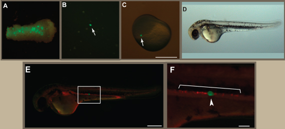 Transplantation of a single PGC from a sturgeon embryo into a goldfish embryo. (A) A removed fragment containing PGCs from sturgeon embryo at stage 26. (B) Dissociated cells from the fragment. Arrow indicates a GFP-labeled PGC. (C) A goldfish embryo at the blastula stage showing the location of an isolated single PGC following transplantation (arrow). (D) A PGC transplanted chimera at 3-dpf. (E) Fluorescence view of (D). (F) The magnified image of the box in (E). RFP-labeled cells indicate endogenous goldfish PGCs and GFP-labeled cell indicates the transplanted sturgeon PGC. Note that the sturgeon PGC localized together with goldfish PGCs at the gonadal region. Scale bars in (C) and (E) indicate 500 µm; the scale bar in (F) indicates 100 µm. doi:10.1371/journal.pone.0086861.g005
| Press Release | ||
|---|---|---|
| Key Points |
* The primordial germ cells (PGCs) of a sturgeon, which become eggs or sperm, have been found to have the same mechanism as that of a frog, and they emerge during embryonic development. * When transplanted into a goldfish embryo, which develops differently from the embryo of a sturgeon, the PGCs migrated to its gonads. * These findings show that the development and migration of the cells to become eggs or sperm have similarities among species. |
|
| Overview |
The over-fishing of caviar, one of the world’s three great delicacies, has caused a significant decrease in their number, resulting in the need for the conservation of wild sturgeon. Sturgeon are regarded as living fossils, and are important in order to solve the mysteries of evolution. Today, technology has been developed to allow a sturgeon’s gametes (eggs or sperm) to be formed in other species so as to revive these endangered fish. To achieve this goal, we must first learn how the primordial germ cells become gametes. A sturgeon is categorized as a fish; however, the process of its embryonic development is very similar to that of frogs. Observation of the colored part of the embryo (a fertilized egg) show that sturgeon PGCs emerged in the same mechanism as those of a frog. What’s more, when these PGCs were transplanted into a goldfish embryo, which develop a different cleavage pattern from the embryo of a sturgeon, they were found to have the ability to migrate to the goldfish’s gonads. These results indicate that the development and migration of cells that are potential eggs or sperm have similarities among species. |
|
| Inquiries |
Etsuro Yamaha, Professor, Nanae Fresh-Water Laboratory, Aquatic Research Station (Jpn link); Field Science Center for Northern Biosphere TEL: +81-138-65-2344 FAX: +81-138-65-2239 E-mail: eyamaha@fsc.hokudai.ac.jp |
|
|
Japanese Link |
キャビアの元となる細胞の由来~キャビアとなる細胞はチョウザメ受精卵からどのように出現するのか?~ | |
| Publications | PLoS One (2014.2.5) | |