Neural Mechanisms for Integrating Retinal Images Before and After Saccadic Eye Movement and Maintaining a Stable and Continuous Visual World
Research Press Release | March 24, 2016
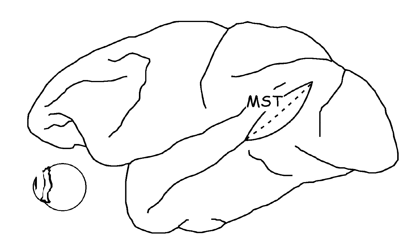
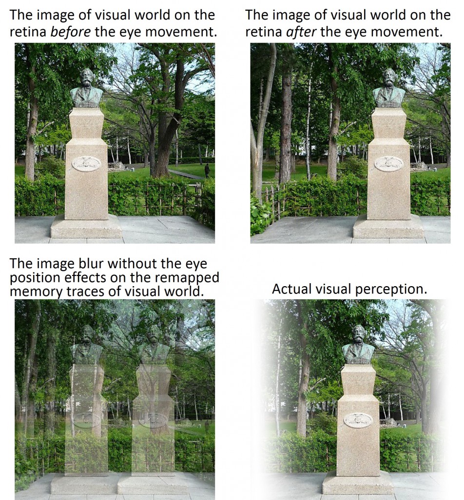
Figure 2. The images of visual world before and after the eye movements.
|
Outline |
Our eyes move rapidly and with an extremely high frequency of several times per second. Yet we are able to perceive the visual world as stable and continuous despite our eye movements, unlike a camera’s blur. How does our brain deal with the blurry retinal images resulting from self-eye-movements? In order to understand the neural mechanisms of the visual stability irrespective of eye movements, we recorded and analyzed neural activity in the MST area, which is part of the posterior parietal cortex of macaque monkeys (Figure 1). So far we have clarified that the MST area contributes to compensate for the retinal image motion induced by smooth pursuit eye movements (Inaba et al., 2007, J Neurophysiol, etc.), and also to recall visual information, that have pre-existed before saccadic eye movement, after the eye movement (Inaba and Kawano, 2014, PNAS). However, even if visual information prior to eye movement can be recalled after a saccade, the visual image before and after the eye movement would be displaced by the amount that the eye moved, so one would expect that a gap would occur in the visual world before and after the saccade (Figure 2). In the present study we investigated the post-saccadic eye position effects on the neuronal responses to the visual memory traces which is recalled after the saccade, and clarified that the MST neurons simultaneously integrate the information of the post-saccadic eye position with the pre-saccadic visual information that is recalled after eye movement. This result suggests that the MST neurons integrate the two displaced retinal images before and after saccadic eye movement by using eye position information before and after the eye movement, and in this way contribute to maintaining a stable and continuous visual world. |
|
| Inquiries |
Assistant Professor Naoko INABA Systems Neuroscience Laboratory, Department of Physiology, Hokkaido University Graduate School of Medicine ninaba[at]med.hokudai.ac.jp |
|
|
Japanese Link |
眼の動きの前後の網膜像を統合し 滑らかな視界を維持する脳の仕組みを解明 (02.25.2016) | |
|
Publications |
Eye position effects on the remapped memory trace of visual motion in cortical area MST, Scientific Reports (02.24.2016) | |