Hokkaido University Leads the World in Radiation Therapy as a Result of Industry-Academia Cooperation
Research Press Release | July 28, 2015
|
Proton Beam Therapy System Jointly Developed with Hitachi, Ltd. will be Installed at a World Leading Hospital |
||
|---|---|---|
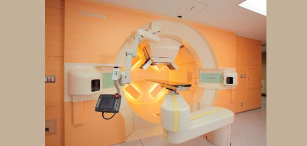
| Key Points | ・It has been decided that world-leading proton beam1 therapy system jointly developed by Hokkaido University (President Keizo Yamaguchi) and Hitachi, Ltd. (Representative Executive Officer and President and COO Toshiaki Higashihara) under the FIRST Program2, will be installed at Sibley Memorial Hospital3 (Washington, D.C., U.S.A.), which is affiliated with Johns Hopkins Medicine, one of the leading hospitals in the world.
・Research and development on the proton beam therapy system combined real-time tumor-tracking radiation therapy that is the intellectual property of Hokkaido University with Spot-scanning Technique5 of Hitachi, will be a driving force for radiation therapy for the world. |
|
| Background & Overview | Radiation therapy, one of the three pillars of cancer therapy, is a “therapy that is easy on the patient” because it is painless. For this reason, it is hoped that it will become the leading cancer therapy of the future. X-rays are conventionally used for therapeutic radiation, but therapy equipment that uses “proton beams” has recently begun to see wider use, especially in developed countries, because it has less effect than X-rays on the surrounding normal tissue, which is the cause of side effects.
“Real-time tumor-tracking radiation therapy” which was developed in the 1990s, can accurately irradiate lesions in the trunk of the body that move due to respiratory movement, and is now being used in many X-ray therapy facilities as an effective technique for reducing the adverse effects on healthy tissue. There were many technical problems with the incorporation of this technology into proton beam therapy equipment, but the industry-academia cooperation system of Hitachi and the research team led by Professor Hiroki Shirato of Hokkaido University Graduate School of Medicine was able to overcome these problems. Based on the results, Hitachi has put new proton beam therapy equipment on the market, and this result of industry-academia cooperation has begun to see wider use throughout the world and is entering the stage where it is leading progress in radiation therapy for the world. The new proton beam therapy system, which is the result of joint research and development of Hokkaido University and Hitachi under the FIRST Program supported by the Cabinet Office, will be installed at Sibley Memorial Hospital (Washington, D.C., U.S.A.), affiliated with Johns Hopkins Medicine, one of the leading hospitals of the world. This enhanced proton beam therapy system, which was first presented to the world by Professor Hiroki Shirato of Hokkaido University, combines real-time tumor-tracking radiation therapy that is Hokkaido University’s intellectual property with Hitachi’s spot-scanning technique. By combining these two types of irradiation technique, it became possible to irradiate with a proton beam with high precision even on a tumor whose position has changed due to respiration, which greatly reduced the irradiation of healthy tissue. The accelerator, an important component of the proton beam therapy system made based on the Hokkaido University model has also been installed at the world-leading Mayo Clinic in the U.S.A—one leading hospital after another is introducing the technology of Hokkaido University and Hitachi— and within Japan, therapy has already started at the Hokkaido University Hospital. Characteristics of the proton beam therapy system that make it possible to irradiate all types of tumors with high precision include miniaturization of the equipment as the result of being specialized for spot-scanning therapy (pharmaceutical approval obtained February 2014), as well as the incorporation of real-time tumor-tracking radiation therapy (approval obtained October 2014), and the addition of a gantry-mounted cone beam CT6 (approval obtained March 2015). These next-generation improvements greatly influenced the selection of the system and will drive proton beam therapy in the future. Hokkaido University is forming an alliance within the University to strongly promote medical-engineering collaborative systems. In addition to this FIRST Program, the alliance includes the Graduate School of Medicine, the Faculty of Engineering, Hokkaido University Hospital, the Hokkaido Organization for Translational Research, GI-CoRE (Global Institution for Collaborative Research and Education)—which reports directly to the President and supports international collaborative research and education—and the Institute for the Promotion of Business-Regional Collaboration . This will lead to more results in international industry-academia cooperation. Encouraged by the current market response, Hokkaido University is further promoting research and education related to medicine, science, and engineering, and will continue to push forward with joint industry-academia research in which industry and academia become united, and will promote radiation therapy throughout the world with technology developed in Japan. |
|
| Terms: |
1 Proton Beam: A proton beam has a physical characteristic called a Bragg peak, in which most of the energy is emitted near the end of the arrival depth. Since a proton beam emits most of its energy near the lesion, an important characteristic is that it does not reach behind the lesion. X-rays, on the other hand, have the property that the energy is a maximum at a fixed depth from the body surface, and after that the X-ray gradually decreases, and as a result of this property a fixed amount of radiation reaches the normal tissue behind the lesion. 2 FIRST Program: The “Funding Program for World-Leading Innovative R&D on Science and Technology”, is a large-scale research support system set up as a science and technology policy. It began accepting applications in 2009, and out of 565 applications from all over Japan, in March, 2010, the Council for Science and Technology determined thirty “core researchers and research projects” that will support the future of Japanese science and technology. From Hokkaido University, the “Advanced Radiation Therapy Project -Real-time Tumor-tracking with Molecular Imaging Technique-”, of Professor Hiroki Shirato of the Graduate School of Medicine, was selected. It was the only program selected in the field of radiation therapy, and received attention both within and outside Japan as a project that would lead the future development of radiation therapy and cancer therapy technology. 3 Sibley Memorial Hospital (Washington, D.C.): A hospital that is affiliated with Johns Hopkins Medical. It always receives a top rating in the U.S. hospital rankings. It opened in 1890. 4 Real-time Tumor-tracking Radiation Therapy: Real-time tumor-tracking radiation therapy is conducted by implanting 2 mm gold markers near the tumor. It first uses CT equipment to assess the surrounding area between the markers and the center of the tumor via X-ray fluoroscopy from two directions to automatically extract the gold marker on the fluoroscopic image with pattern recognition technology. It periodically re-calculates the spatial positions. After this, irradiation is performed only if the gold markers are within ranges of several mm from the planned position. By doing this at high speed, it is possible to perform highly accurate irradiation even on cancers that change their position within the body due to respiration. Compared to a method that irradiates the entire range that the cancer moves, the irradiated volume is reduced to 1/2 to 1/4, making it possible to greatly reduce the irradiation of healthy tissue. 5 Spot-Scanning Technique: Spot-scanning therapy does not diffuse the proton beam that irradiates the tumor as in previous methods, but rather is a technology that irradiates with a proton beam by taking advantage of its thinness to repeatedly alternate between a high speed of irradiation and temporary pauses while successively changing its position. Therefore, even for tumors that have complicated shapes, it is possible to irradiate with the proton beam with high accuracy matching the required shape, while keeping the adverse effect on healthytissue to a minimum. 6 Gantry-Mounted Cone Beam CT: This is a technology that uses an X-ray tube and an imaging device mounted on a gantry (a large circular stand) in order to obtain a cone beam (an X-ray beam in the shape of a cone) CT image, and that makes it possible to view soft tissue within the body. This is used to determine highly accurate target positions. |
|
| Inquiries |
Institute for the Promotion of Business-Regional Collaboration, Hokkaido University TEL: +81-11-706-9561 FAX: +81-11-706-9550 E-mail:jigyo@mcip.hokudai.ac.jp |
|
|
Japanese Link |
北海道大学の産学連携成果が世界の放射線治療をリード~日立製作所との共同開発による陽子線治療装置が世界トップクラスの病院に導入決定~ (2015.7.10) | |