Spotlight on Research: Going nano to enhance drug delivery systems and single cell analytics
Research Highlight | December 06, 2019
Nanotechnology is a buzz word. With nanomaterials sweeping nations worldwide, it seems like everyone is looking for ways to make new tech smaller in size. You likely have heard how nanotechnology is revolutionizing the field of electronics, but did you know it’s also working its magic in other fields?
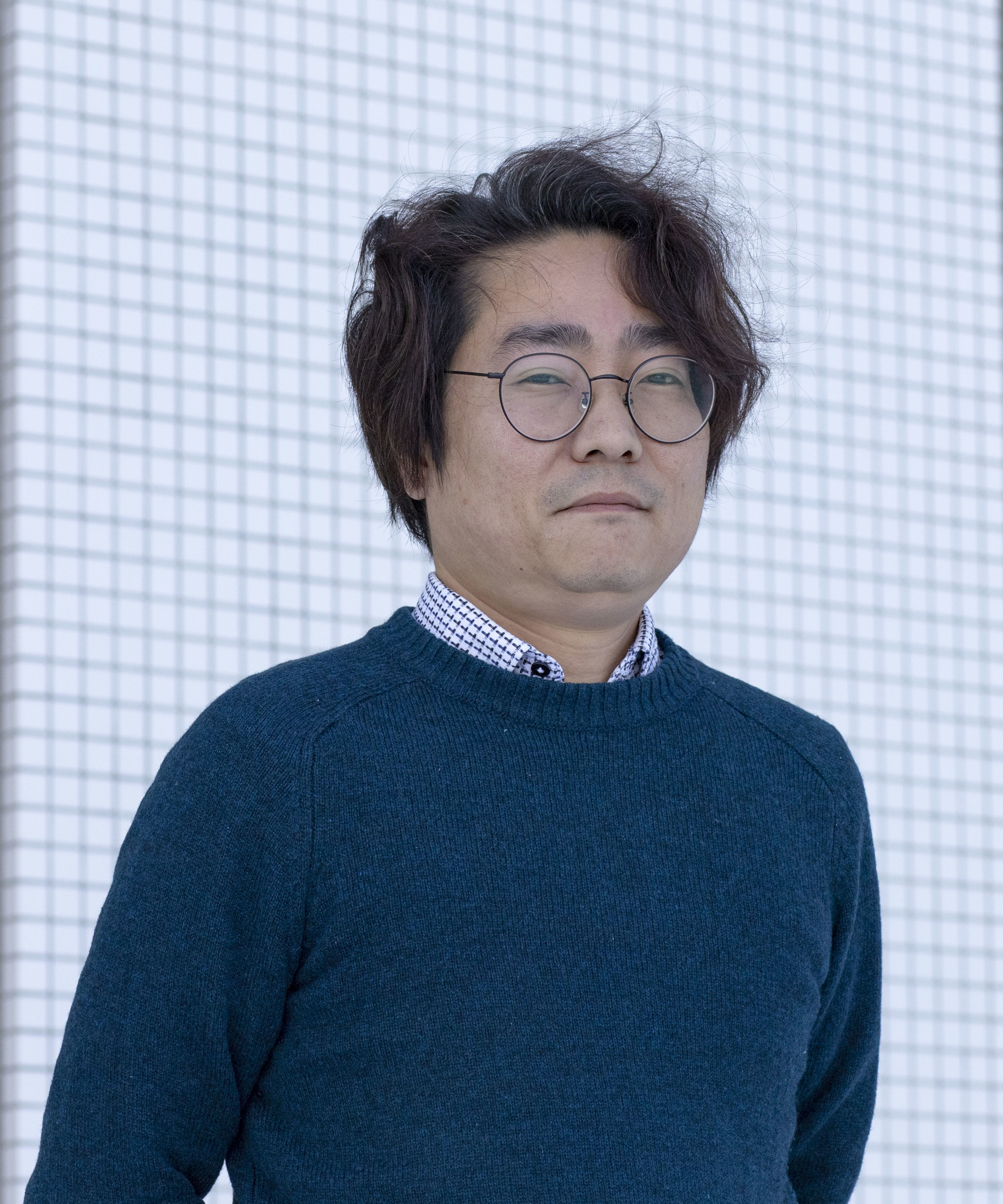
Professor Hiroshi Uji-i with Hokkaido University’s Research Institute for Electronic Science has developed a new optical fiber made of metal to more effectively deliver anti-cancer drugs in our bodies and understand what exactly is going on in each individual cell.
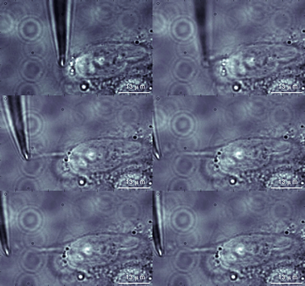
The metallic nanowires, which are made in Professor Uji-i’s laboratory, are so tiny, 100 nanometers thin— that’s 0.1 micrometers—that a slightly larger wire needs to be placed over it to control its movements.
Similar to optical fiber, light is shot through one side and sent to the other. Except, instead of sending information to the other side, the light is scattered and returns in the same direction it came. In doing so, Professor Uji-i and his laboratory receive spectroscopic information, essentially light emission and radiation information, from any location within a cell. This information can tell us how and when a drug enters a cell, and the ways they interact with their surroundings.
“We use a technique called Raman spectroscopy,” Professor Uji-i explained. “Traditionally, surface enhanced Raman spectroscopy produces too much background noise. It also generates heat, so you may burn or even kill the cell. But, by making the nanowires out of silver or gold, the quality of information is automatically enhanced, expanding what has been possible for us up until this point.”
Using this method, Professor Uji-i studies how anti-cancer drugs are delivered to the tumor site, how they get into the cancer cells, and how they interact with the proteins and DNA inside.
“At the moment, most cancer treatments use drugs that also kill healthy cells,” he continued. “This is why people suffer from serious side effects. The drugs circulate throughout the entire body. Some go to the tumor and some don’t. If we can know how the drugs interact on a molecular level, we can design safer and more efficient anti-cancer drugs and delivery systems.”
Professor Uji-i started out as a microscopist and has always been interested in studying small things that the eye cannot see. He then moved to Belgium at the University of Leuven to specialize in single molecular spectroscopy, studying properties at the sub-micron level. After moving back to Japan 4 years ago, he wanted to keep pushing the boundaries of how small we can actually go, and that’s when he took his research to the nanoscale.
“There is no molecular view of this research. There is almost none, and that is scary,” he pointed out. “The information we have on how effective a drug is working, for example, is averaged, not on the individual cell level, and I want to change this.”
The initial results of the new drug delivery system were first published earlier this year. Professor Uji-i plans to publish within the year further findings elucidating interactions at the molecular level within the cell as a result of the nanowire techniques he developed.
The nanoscopy and nanowire research taking place at Professor Uji-i’s laboratory can be used for a variety of purposes. Members of his laboratory are looking into ways to map DNA and selectively detect molecules, for example. All of these research topics will of course continue to push forward the field, but what Professor Uji-i is particularly interested in is its potential uses for iPS cell technology.
iPS cells, or induced pluripotent stem cells, are genetically modified cells that can be turned into any type of cell, a muscle cell or even a bone cell. Ongoing research is particularly prevalent in Japan, with clinical trials already starting to use the technology.
In 2015 Professor Uji-i was contacted by leading iPS researcher Prof. M. Sampaolesi of the University of Leuven to investigate an anomaly in some iPS cell behaviors. After a while, some of the modified cells had mutated into a completely different cell type, triggering the surrounding cells to also change.
In response, Professor Uji-i is hoping to develop a nanowire to track how cells change over time as well as what he calls a “nano-syringe” to help improve and ensure iPS technology is safe to use.
“I believe iPS technology may be used on a daily basis in the future for medical treatments. We may end up with a lot of modified genes in our body,” said Professor Uji-i. “The danger is that we don’t know how these modified genes develop in the body or whether they would pass to your offspring. My dream is to develop a new method to track a single cell, a single cell analytical method.”
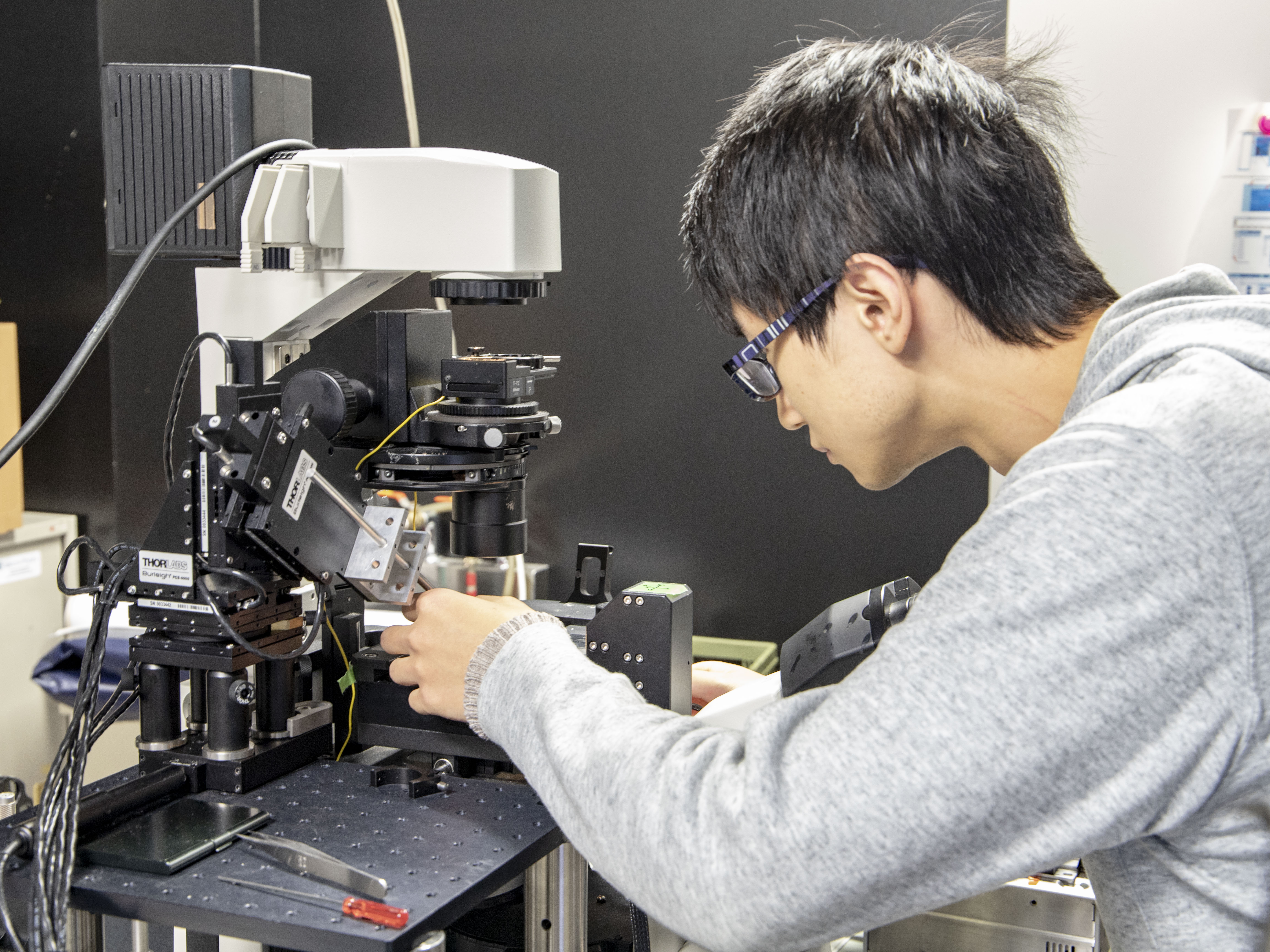
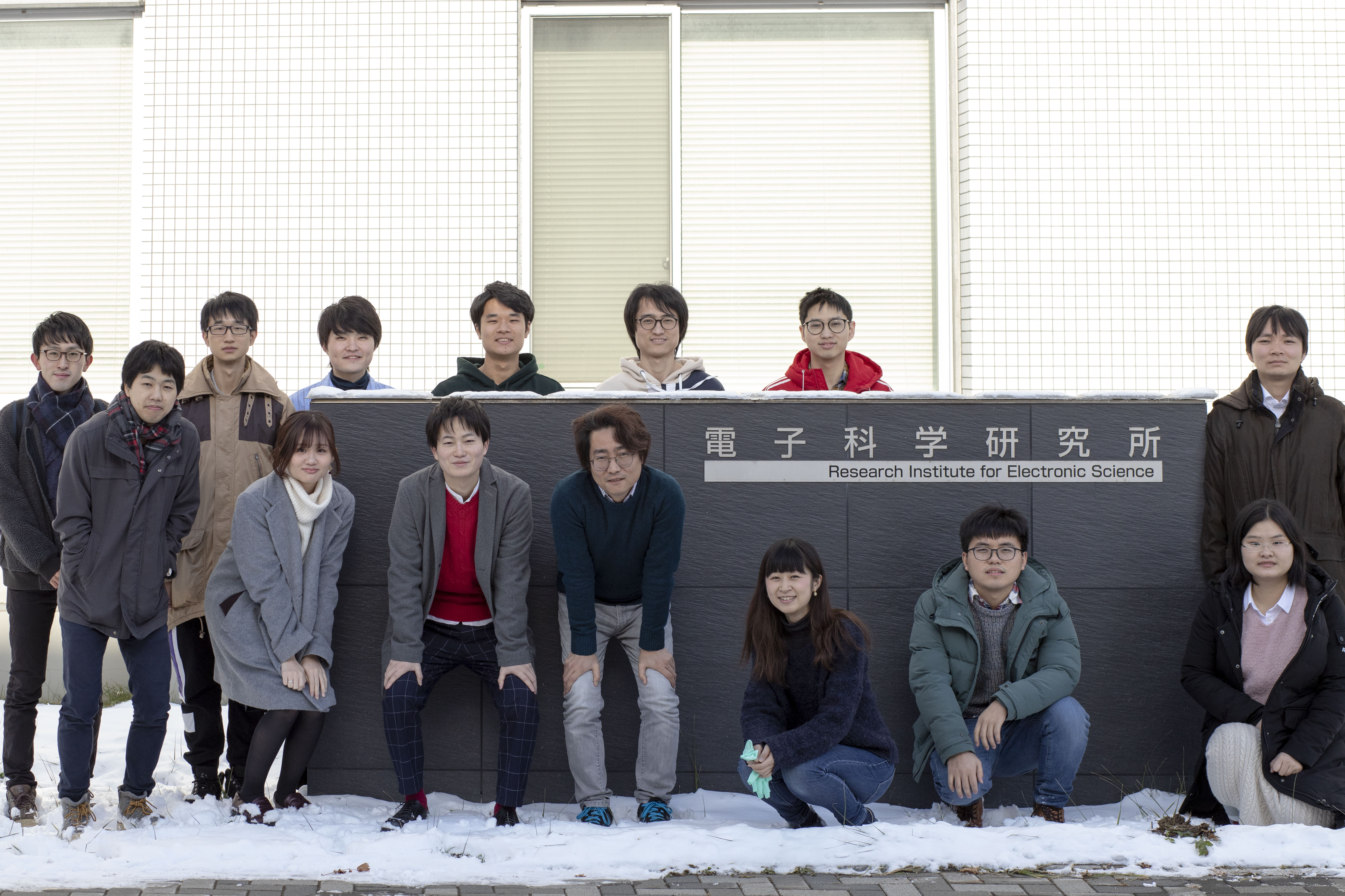
Professor Uji-i and his laboratory’s research are made possible because the members are of diverse scientific backgrounds, leading to transdisciplinary collaborations and projects. Any student or faculty interested in this research is encouraged to reach out to the Uji-i Laboratory at any time.
Researcher details:
Professor Hiroshi Uji-i
Research Institute for Electronic Science
Hokkaido University
hiroshi.ujii@es.hokudai.ac.jp
Author: Dr. Katrina-Kay Alaimo


