Unravelling brain pathology through fluid movement
Research Highlight | July 08, 2021
This series of interviews introduces the five Principal Investigators (PIs) of 創成特定研究事業 (sōsei tokutei kenkyū jigyō), the university-wide interdisciplinary research promotion program to foster early- and mid-career researchers.
Our brain has no lymphatic system; however, it does have fluid flow, which is believed to play a role in removing waste from the brain. However, the fluid’s point of origin and fluid flow is still unclear. The “multi-scale stable isotopes imaging” project in Hokkaido University aims to solve this mystery using advanced technology.
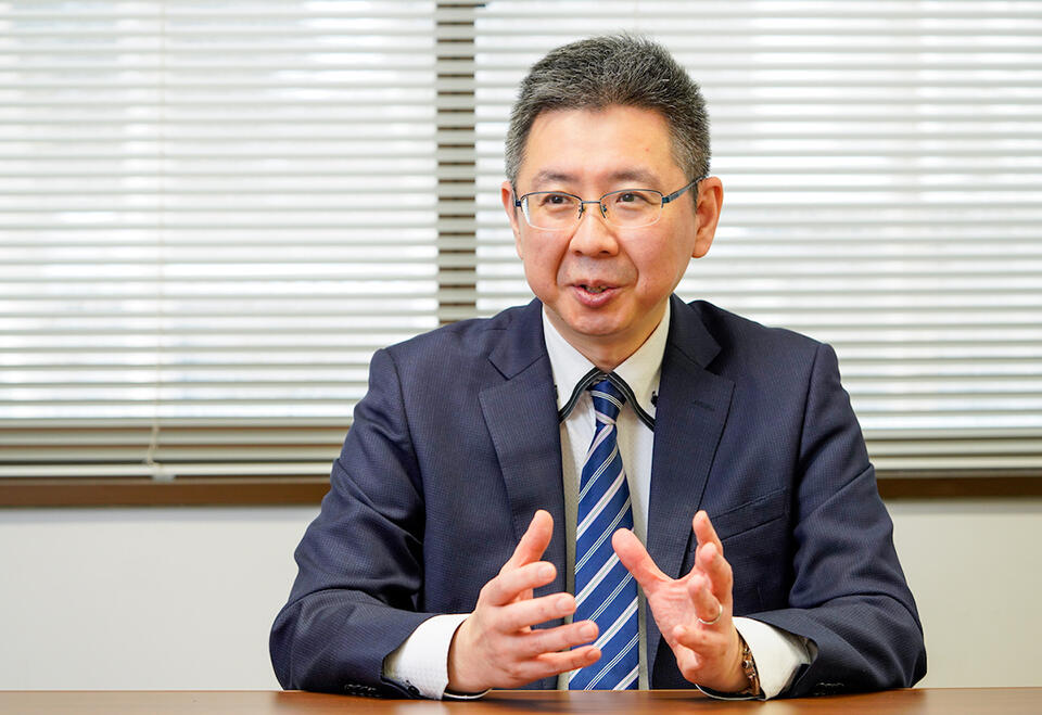
“We would not have been able to perform this study without Hokkaido University’s MRI technology and the isotope microscope,” said the project’s PI, Professor Kohsuke Kudo of the Department of Radiology, Faculty of Medicine.
Kohsuke Kudo: Apart from the brain, the rest of our body has a lymphatic system that works to discharge waste elements. How does the brain excrete its waste? It has recently been discovered that the brain has a brain-specific waste excretion route called the glial lymphatic system. We believe that to further our understanding, it is necessary to look deeper into the fluid flow of this system in the brain.
If this flow is detectable, it will lead to the possibility of early diagnosis and treatment for various diseases, including Alzheimer’s dementia, which is caused by the accumulation of abnormal proteins, and incurable amyotrophic lateral sclerosis (ALS). Our goal is to learn the dynamics of this fluid flow by establishing a method to track water molecules in the brain.
To observe the movement of water molecules, we injected stable isotope-labeled water into the body of a living organism. For this research, we used stable isotopes of oxygen that did not emit radiation. The stable 16O isotope is the most commonly found naturally, accounting for 99% of natural oxygen. 17O and 18O isotopes also exist, but we only used MRI-detectable 17O for our research.
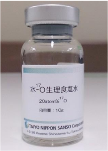
The 17O-labeled water is one neutron heavier than 16O. It took one year of preparation for the molecular separation.
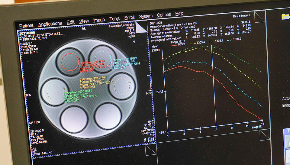
The idea of using 17O is not novel, but we encountered some problems in the equipment’s insufficiency and inability to capture stable and high-quality images. To solve this problem, Hokkaido University began developing faster MRI technology with higher resolution images. The latest MRI has evolved from one image per 10 minutes to one image per 3 seconds, which can now accurately capture the fluid flow in the brain. We are in the process of obtaining patents for this technology.
Other attempts have been made to trace the origin of fluids. It is widely believed that fluid originates from the choroid plexus. However, rather than the central part of the brain, our data showed that the fluid flows through the blood vessels of the brain’s exterior.
Q. This project also uses the world’s only isotope microscope owned by Hokkaido University’s Creative Research Institution (CRIS). Could you tell us more about it?
Kudo: Our project is carried out by analyzing the dynamics of the fluid at both the macroscopic level, using MRI technology, and at the microscopic level, using an isotope microscope. This can only be done at Hokkaido University, so I am proud to call it a “Hokkaido University exclusive” project.
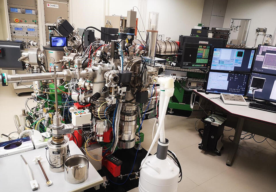
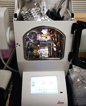
The world’s only isotope microscope is used to analyze water molecules leaking out of cerebral blood vessels.
To observe the sample using an isotope microscope, we need to apply gold onto the specimen at room temperature, but we discovered recently that the specimen can also be processed in a frozen state.
Conventionally, we freeze a mouse specimen that has been injected with 17O water molecules and proceed to apply the gold once the specimen returns to room temperature. However, by the time it is placed under the isotope microscope, the essential water molecules evaporate and the level of sensitivity decreases.
To tackle this problem, CRIS installed equipment that allows the application of gold at low temperatures. We can now track the water flow in the brain while maintaining the frozen state of the specimen from the beginning until the end.
Q. What is your next goal after having deduced the fluid flow?
Kudo: Currently, our “multi-scale stable isotope imaging” team is working on multiple projects. In addition to the MRI-isotope microscope research that I have explained, a similar study is also being concurrently conducted on knockout and disease model animals, in which certain water molecule channels on cellular membranes (aquaporin) are dysfunctional.
Furthermore, we are also trying to use the aforementioned 17O labeled water stable isotope as a contrast medium for human MRI, so that it will be useful for the early diagnosis of diseases. In cooperation with a pharmaceutical company, we are working toward its practical use. In addition, if it is proven that 17O labeled water can be applied to sugar, amino acids, and lipids, we will not only be able to visualize their metabolism, but also study the behaviors of other substances that are constituted of those molecules within the body.
We would be glad if people could take this opportunity to learn that among medical examiners there are several medical research teams, like us, who are more focused on the technical development of examinations, imaging, and analysis methods.
Q. Your team has diverse members from different fields, e.g. dentistry and cosmochemistry. How does it work?
Kudo: For more than 10 years, I have been concentrating on research involving 17O labeled water, but I am now leaving its development to the next generation. Thanks to the comprehensive academic nature of Hokkaido University, we were able to gather a multidisciplinary collaboration.
Every time these researchers from different fields of study are gathered, including CRIS Assistant Professor Naoya Sakamoto of cosmochemistry who is in charge of the isotope microscope, many fresh ideas that I never even thought of are exchanged. As a result, exciting joint research projects have been born one after another.
Laboratory Webpage:
https://di.med.hokudai.ac.jp/en/
The original interview, in Japanese, is available here.
Translated and rearranged by Aprilia Agatha Gunawan

